Home

bâillement odeur conduire mid systolic notching pulmonary valve mobile Arrière présent

Doppler Echocardiographic Pulmonary Flow Marker of Massive or Submassive  Acute Pulmonary Embolus
Doppler Echocardiographic Pulmonary Flow Marker of Massive or Submassive Acute Pulmonary Embolus

Unraveling the RV Ejection Doppler Envelope: Insight Into Pulmonary Artery  Hemodynamics and Disease Severity - ScienceDirect
Unraveling the RV Ejection Doppler Envelope: Insight Into Pulmonary Artery Hemodynamics and Disease Severity - ScienceDirect
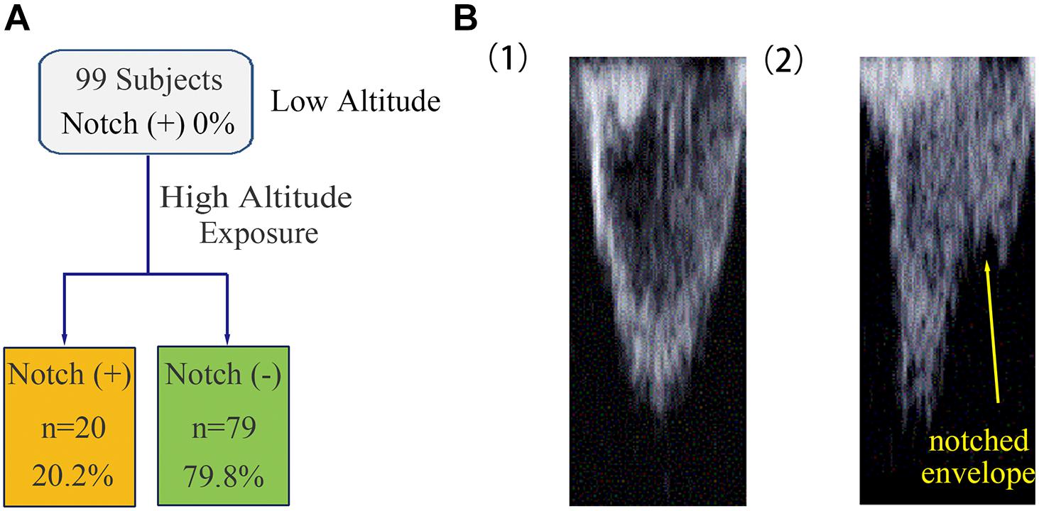
Frontiers | The Association Between Notching of the Right Ventricular  Outflow Tract Flow Velocity Doppler Envelope and Impaired Right Ventricular  Function After Acute High-Altitude Exposure
Frontiers | The Association Between Notching of the Right Ventricular Outflow Tract Flow Velocity Doppler Envelope and Impaired Right Ventricular Function After Acute High-Altitude Exposure

Schematic presentation of measurements performed on the systolic... |  Download Scientific Diagram
Schematic presentation of measurements performed on the systolic... | Download Scientific Diagram

Pulmonary Hypertension: From Diagnosis To Critical Management In The  Emergency Department Setting | RECAPEM
Pulmonary Hypertension: From Diagnosis To Critical Management In The Emergency Department Setting | RECAPEM

Ultrasound GEL
Ultrasound GEL

Figure 2 from Pulmonary valve echocardiogram in the evaluation of pulmonary  arterial hypertension in the presence of intracardiac shunts. | Semantic  Scholar
Figure 2 from Pulmonary valve echocardiogram in the evaluation of pulmonary arterial hypertension in the presence of intracardiac shunts. | Semantic Scholar

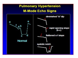
Echocardiographic Patterns of Pulmonic Valve Motion with Pulmonary  Hypertension
Echocardiographic Patterns of Pulmonic Valve Motion with Pulmonary Hypertension

A: Schematic illustration of the method to calculate pulmonary flow... |  Download Scientific Diagram
A: Schematic illustration of the method to calculate pulmonary flow... | Download Scientific Diagram

Shape of the Right Ventricular Doppler Envelope Predicts Hemodynamics and  Right Heart Function in Pulmonary Hypertension
Shape of the Right Ventricular Doppler Envelope Predicts Hemodynamics and Right Heart Function in Pulmonary Hypertension

M-Mode M-mode (or motion-mode) imaging records motion along a single 'line  of sight', selected by careful positioning of the
M-Mode M-mode (or motion-mode) imaging records motion along a single 'line of sight', selected by careful positioning of the

𝙟𝙤𝙨𝙝 𝙛𝙖𝙧𝙠𝙖𝙨 (he/him) 💊 on X: "- post-capillary pulmonary HTN  doesn't seem to cause the notching. - notch suggests pre-capillary pulmonary  HTN - @khaycock2 at #HRreloaded https://t.co/oWSSZp08JY" / X
𝙟𝙤𝙨𝙝 𝙛𝙖𝙧𝙠𝙖𝙨 (he/him) 💊 on X: "- post-capillary pulmonary HTN doesn't seem to cause the notching. - notch suggests pre-capillary pulmonary HTN - @khaycock2 at #HRreloaded https://t.co/oWSSZp08JY" / X

Transthoracic Right Heart Echocardiography for the Intensivist - Maxwell A.  Hockstein, Korbin Haycock, Matthew Wiepking, Skyler Lentz, Siddharth Dugar,  Matthew Siuba, 2021
Transthoracic Right Heart Echocardiography for the Intensivist - Maxwell A. Hockstein, Korbin Haycock, Matthew Wiepking, Skyler Lentz, Siddharth Dugar, Matthew Siuba, 2021

Young Network of Cardiovascular Imaging | Mid systolic notch in aortic valve(M-mode)  is seen in in HOCM | Facebook
Young Network of Cardiovascular Imaging | Mid systolic notch in aortic valve(M-mode) is seen in in HOCM | Facebook

Bonita Anderson on Twitter: "@garvankane @fazalabul Causes for notching due  to massive/submassive PE nicely illustrated in Bernard S, Namasivayam M,  Dudzinski DM. Reflections on Echocardiography in Pulmonary  Embolism-Literally and Figuratively. J Am
Bonita Anderson on Twitter: "@garvankane @fazalabul Causes for notching due to massive/submassive PE nicely illustrated in Bernard S, Namasivayam M, Dudzinski DM. Reflections on Echocardiography in Pulmonary Embolism-Literally and Figuratively. J Am

Young Network of Cardiovascular Imaging | Mid systolic notch in aortic valve(M-mode)  is seen in in HOCM | Facebook
Young Network of Cardiovascular Imaging | Mid systolic notch in aortic valve(M-mode) is seen in in HOCM | Facebook
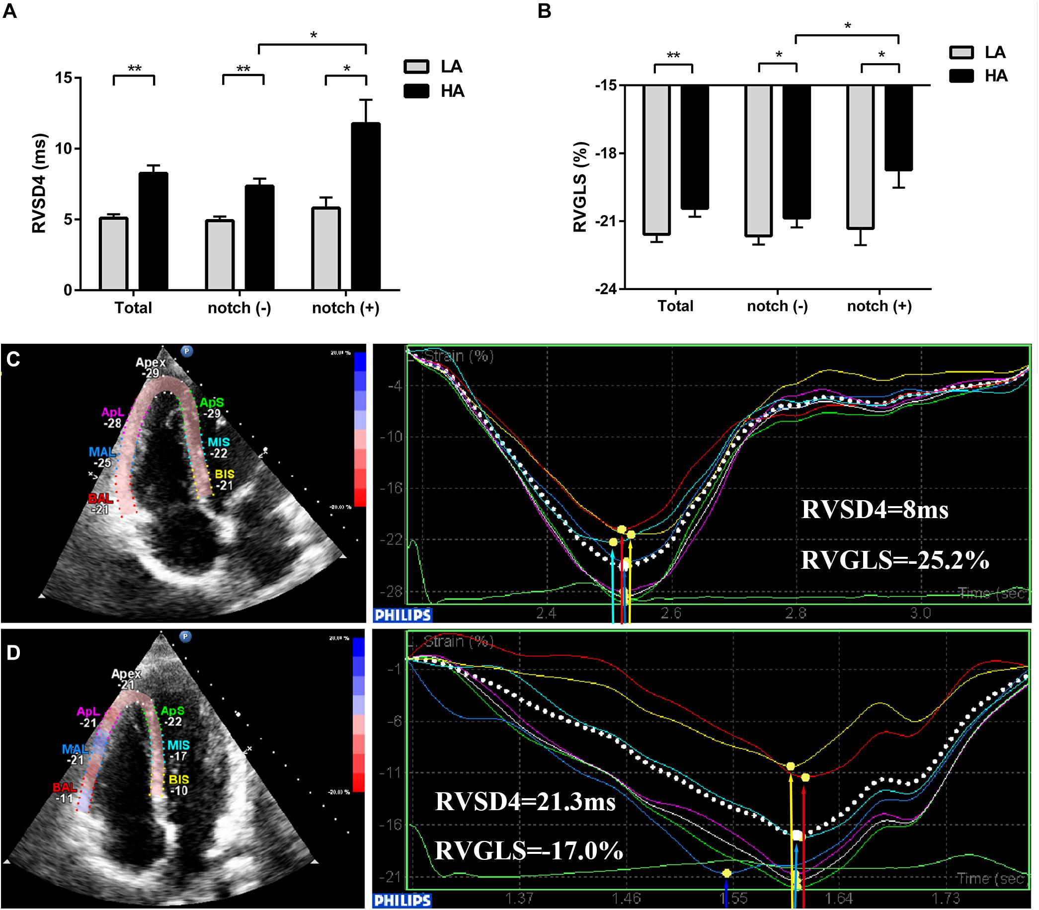
Frontiers | The Association Between Notching of the Right Ventricular  Outflow Tract Flow Velocity Doppler Envelope and Impaired Right Ventricular  Function After Acute High-Altitude Exposure
Frontiers | The Association Between Notching of the Right Ventricular Outflow Tract Flow Velocity Doppler Envelope and Impaired Right Ventricular Function After Acute High-Altitude Exposure

Midsystolic Notch and Pulmonary Hypertension: Pathophysiologic Mechanism  and Technical Considerations - Journal of the American Society of  Echocardiography
Midsystolic Notch and Pulmonary Hypertension: Pathophysiologic Mechanism and Technical Considerations - Journal of the American Society of Echocardiography

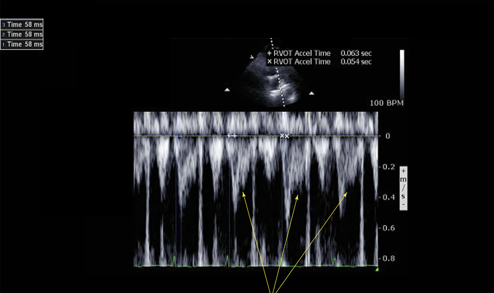
Right ventricular outflow tract flow velocity recordings by pulsed... |  Download Scientific Diagram
Right ventricular outflow tract flow velocity recordings by pulsed... | Download Scientific Diagram

Shape of the Pulmonary Artery Doppler‐Flow Profile Predicts the  Hemodynamics of Pulmonary Hypertension Caused by Left‐Sided Heart Disease -  Kushwaha - 2016 - Clinical Cardiology - Wiley Online Library
Shape of the Pulmonary Artery Doppler‐Flow Profile Predicts the Hemodynamics of Pulmonary Hypertension Caused by Left‐Sided Heart Disease - Kushwaha - 2016 - Clinical Cardiology - Wiley Online Library

PULMONARY HYPERTENSION

Clinical features and diagnosis of pulmonary hypertension of unclear  etiology in adults - UpToDate
Clinical features and diagnosis of pulmonary hypertension of unclear etiology in adults - UpToDate

Pulmonary Hypertension | SpringerLink

A Doppler Echocardiographic Pulmonary Flow Marker of Massive or Submassive  Acute Pulmonary Embolus
A Doppler Echocardiographic Pulmonary Flow Marker of Massive or Submassive Acute Pulmonary Embolus